
OCT 3D Scan
Book an Eye Exam with OCT 3D Scan
What is OCT 3D Scan?
OCT 3D Scan "Optical Coherence Tomography" is an advanced eye scan for people of all ages. Similar to ultrasound, OCT uses light rather than sound waves to illustrate the different layers that make up the back of your eye. The OCT machine captures both a fundus photograph and a cross-sectional scan of the back of the eye at the same time.
The health of your eyes matters to you and it matters to us too, which is why we are offering OCT to all our patients. OCT is a new, completely painless and highly advanced screening system that checks for potentially serious conditions such as glaucoma, diabetes, age-related macular degeneration, vitreous detachments and more.
What can the OCT 3D Scan check for?
Glaucoma

Glaucoma is a condition where the pressure inside the eye is high enough to cause damage to the optic nerve head at the back of the eye. Early diagnosis is key to good prognosis after glaucoma diagnosis. The OCT can pick up glaucoma up to 4 years earlier by mapping the thickness and health of the optic nerve head. Repeated OCT scans allow our Optometrists to detect subtle changes to the Optic Nerve Head which may indicate Glaucoma.
Age Related Macular Degeneration

Macular degeneration causes the gradual breakdown of the macular (the central portion of the eye). OCT can identify this condition and its type (there are two types, wet and dry) and also monitor its progress. Unfortunately the risk of developing macular degeneration increases with age, and it is the most common cause of vision loss in individuals over the age of fifty. Wet macular degeneration causes fluid to leak in the retina and early detection is key to getting treatment.
Diabetic Retinopathy

A patient with diabetest is at risk of a condition called Diabetic Retinopathy where the diabetes affects the eyes. Diabetic retinopathy is a major cause of visual impairment among adults. In the UK, more than two million people have been identified as having diabetes. OCT examination enables early detection, which greatly improves the success rate of treatment. In some cases, diabetes can first be picked up in an eye examination where a referral is made to the GP to ensure the appropriate tests are performed.
Macular Hole
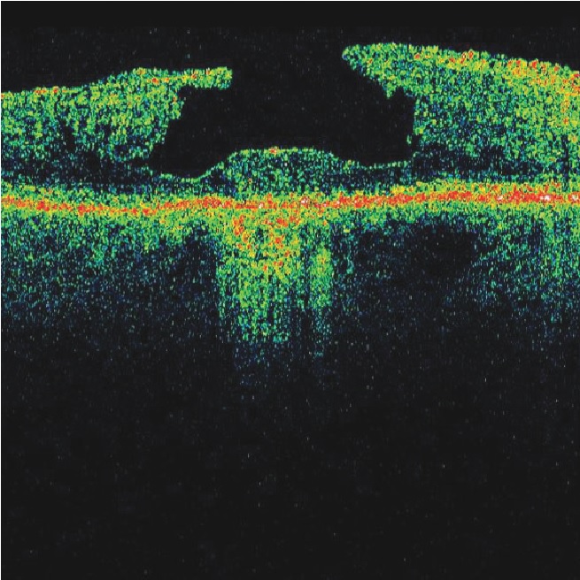
A macular hole is a small hole in the macular – the part of the retina which is responsible for our sharp, detailed, central vision. This is the vision we use when we are looking directly at things, when reading, sewing or using a computer. There are many causes of macular holes. One is caused by vitreous detachment, when the vitreous pulls away from the back of the eye and sometimes it does not ‘let go’ and eventually tears the retina, leaving a hole. Extreme exposure to sunlight (for example staring at the sun during an eclipse) can also cause a macular hole to develop.
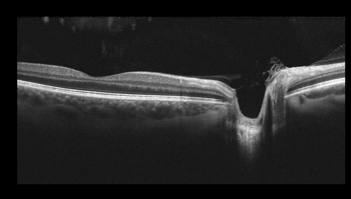
Vitreous Detachment
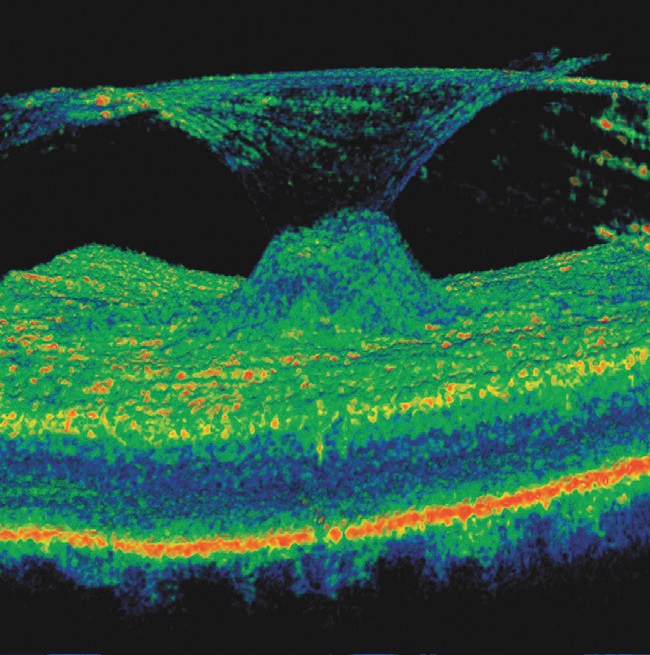
Vitreomacular traction can clearly be diagnosed through OCT providing invaluable information about the current relationship between the vitreous and the retinal surface of the eye. As people get older the vitreous jelly that takes up the space in our eyeball can change. It becomes less firm and can move away from the back of the eye towards the centre, in some cases parts do not detach and cause ‘pulling’ of the retinal surface. The danger of a vitreous detachment is that there is no pain and your eyesight will seem unchanged but the back of your eye may be being damaged
*The OCT 3D Scan is not currently funded by the NHS so private fees apply. Please see our booking page for fees.